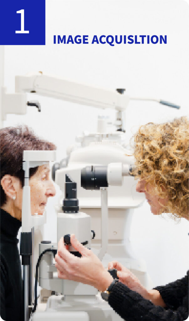
The Pioneering Technology That Will Change the World
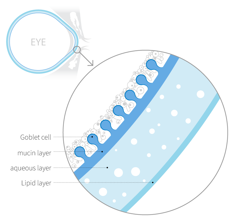
The Tear Film
Dry eye disease occurs due to abnormalities in the tear film and is often accompanied by ocular discomfort and keratoconjunctivitis.The tear film consists of the mucin, aqueous, and lipid layers, and accurate diagnosis of the affected layer is essential for appropriate treatment of dry eye disease.Although methods exist to evaluate each layer of the tear film, there is still a lack of adequate techniques to specifically assess the mucin layer.
“Although several diagnostic modalities are available to assess individual components of the tear film, there remains a significant lack of reliable techniques for specifically evaluating the mucin layer.”

Goblet Cells
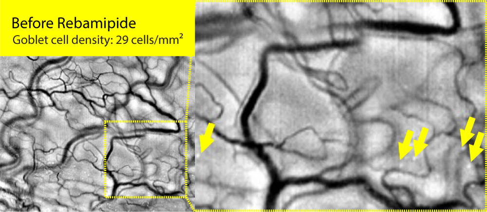
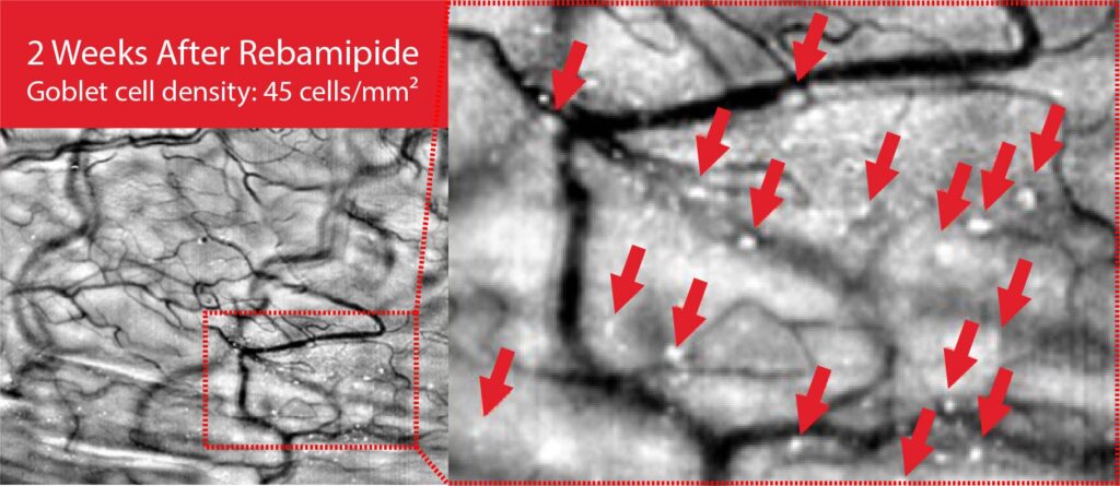
Goblet cells, located in the conjunctival epithelium, secrete the mucin MUC5AC to form the mucin layer of the tear film. They play a key role in maintaining tear film stability and lubricating the ocular surface. When goblet cells are damaged or reduced in number due to inflammation or external stimuli, mucin secretion declines, leading to tear film instability and the onset of ocular surface diseases such as dry eye.
Therefore, goblet cell analysis serves as an important diagnostic biomarker for evaluating the onset, severity, and treatment response of ocular surface disorders, including dry eye disease.
Limitations of Conventional Goblet Cell Examination
One of the most commonly used methods to evaluate goblet cell density and morphological changes is Impression Cytology.
While this technique offers objective assessment of the ocular surface, it is not widely adopted in clinical practice due to the following limitations:
Lack of Standadized Guidelines
The absence of international or national standards leads to inconsistent interpretation of results, as procedures vary across institutions in terms of sampling location, staining method, and evaluation criteria. (e.g., Nelson grading, Tseng method)
Unsuitability for Outpatient Clinical Settings
The procedure requires clinicians to invasively collect cells from the conjunctiva and interpret them under a microscope. Staining and slide preparation demand pathological expertise, and no automated analysis system is available, making the process time-consuming and resource-intensive.
Patient Discomfort and Low Acceptance
As the procedure involves direct contact between the conjunctiva and a thin filter paper, patients may experience discomfort or pain, resulting in low acceptance and compliance.
Disease | Goblet Cell Changes by Disease |
|---|---|
Dry Eye Disease | Tear film instability leads to reduced goblet cell density and impaired mucin secretion. |
Sjögren’s Syndrome | Autoimmune disorder causing decreased tear and saliva secretion, significantly reducing goblet cell density. |
Stevens-Johnson Syndrome | Severe conjunctival damage and extensive loss of goblet cells. |
Allergic Conjunctivitis | Inflammation-induced increase in goblet cell density. |
Conjunctival Intraepithelial Neoplasia (CIN) | Abnormal cell proliferation alters conjunctival structure and disrupts goblet cell morphology. |
Vitamin A Deficiency | Vitamin A deficiency impairs goblet cell function critical for mucin production. |
Chronic Conjunctivitis | Persistent inflammation causes goblet cell loss and dysfunction. |
Chemical Injury | Toxic chemical exposure damages the conjunctiva and destroys goblet cells. |
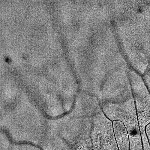
The world’s first imaging-based mucin layer diagnostic solution
We have developed the world’s first imaging technology for conjunctival goblet cells, a core innovation that enables the diagnosis of various ocular diseases, including dry eye disease. Goblet cells serve as definitive biomarkers for dry eye and play a crucial role in maintaining immune tolerance. This breakthrough technology holds potential for expansion into diagnostics for other
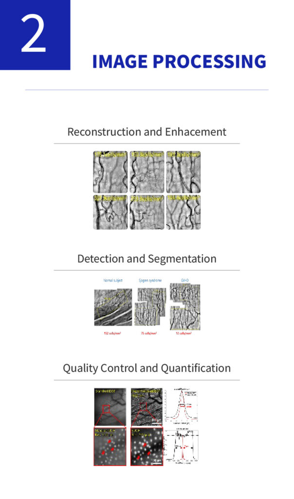
Goblet Cell Examination
Goblet cell examination, which was previously difficult to analyze
Non-invasive, high-precision analytical technology
Accumulated data enables better identification of underlying causes and broadens the potential for guiding treatment strategies
Dry Eye Disease
One of the most prevalent ophthalmic disorders
A multifactorial disease diagnosed by abnormalities in the tear film
Autoimmune Diseases
cludes conditions such as Sjögren’s syndrome and graft-versus-host disease (GVHD)
Characterized by a marked decrease in goblet cell density
Comprehensive Elucidation of the Extended Roles of Goblet Cells
Investigation into the Functional Roles of Goblet Cells within the Mucin Layer and Their Association with Pathological Conditions
: Anticipated acceleration of domain-specific advancements enabled by proprietary technology facilitating direct observation of goblet cells.
Expansion of Moxifloxacin-Based Imaging Techniques
The core moxifloxacin-based cellular imaging platform is anticipated to be applicable not only to ophthalmic diseases but also to a wide range of medical fields such as respiratory and gastrointestinal diseases.
Broadened Deployment of Imaging AI Technologies
Integration of imaging data and AI in the biomedical sector is projected to improve diagnostic accuracy and operational efficiency, thereby expanding potential application fields.
Broadening of Application Fields
Accumulated clinical and learning data will enable broader disease correlation analysis and treatment guidance beyond simple diagnostics.
Specific cell staining and imaging technologies are expected to expand into diverse medical fields.
Improving Diagnostic Efficiency and Identifying Disease Associations through AI Technologies
We are enhancing diagnostic accuracy and efficiency by applying AI-based analysis to high-resolution ocular imaging data. By training machine learning models on micro-level features such as leukocyte movement, goblet cell density, and vascular patterns, we aim to uncover associations with various ocular diseases and broader systemic inflammatory or autoimmune conditions.
To support this, we are increasing investments in software and AI infrastructure, with plans to extend our AI diagnostic applications to areas such as systemic disease evaluation and drug response prediction in the near future.
Artificial Intelligence